What triggers migration? And how can we control and influence it?
Although many parameters do play a role in the answer of such big questions, we focuss on one particular aspect:
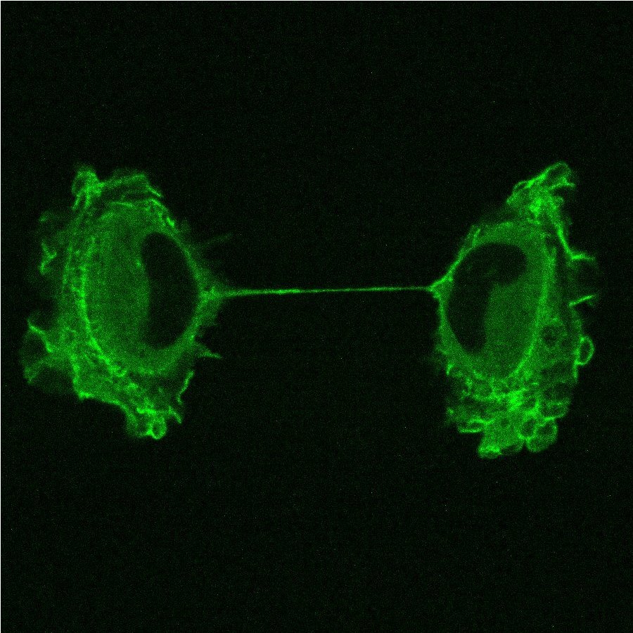
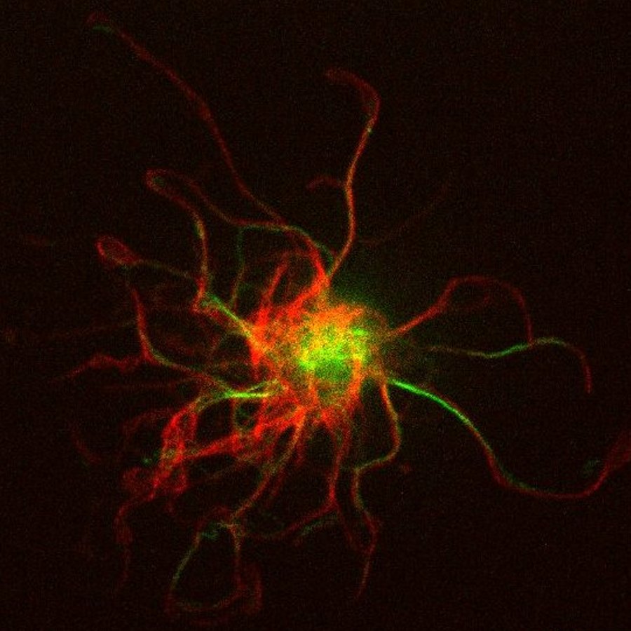
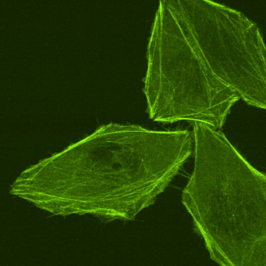
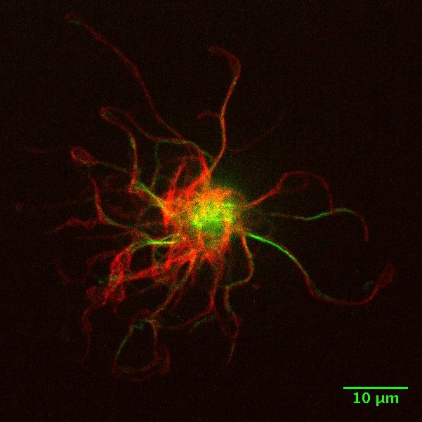
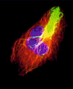
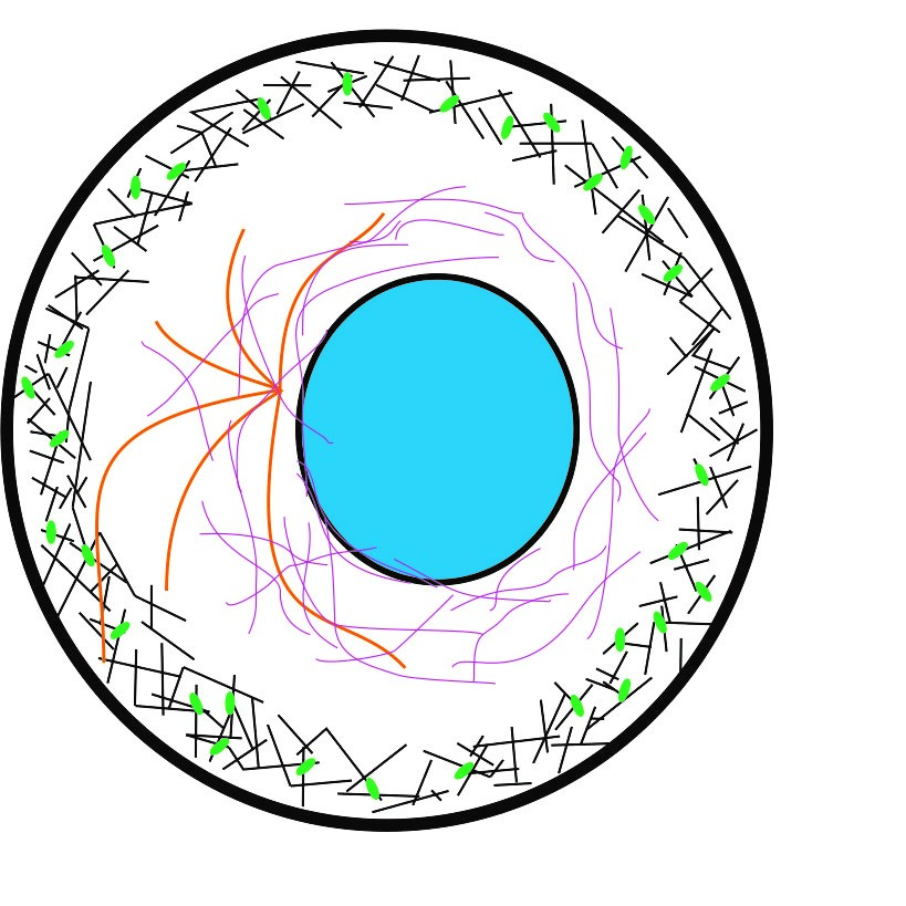
the role of cytoskeletal elements in migrating cells. We investigate the role of actin waves in erratic motion, of intermediate
filaments (especially vimentin) on immune cell migration and the interplay of vimentin with actin. We are also working on the influence of extracellular cytoskeletal
elements on migration.
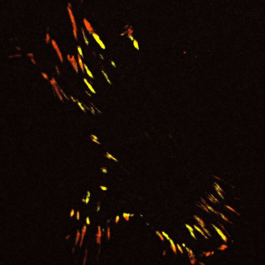
For all migration projects, we are mainly using fluorescent live cell microscopy at different levels of resolution (epi florescence,
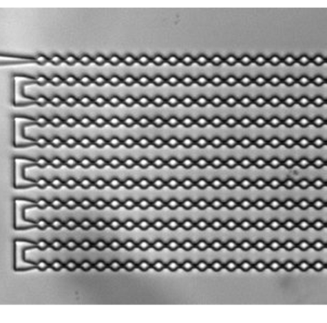
spinning disc confocal microscopy, total internal reflection fluorescence microscopy). For immune cell migration we further need
cellular environments which are confining the cells. We are realizing such environments by creating channels in 1d or by adding precise
roofs on top of 2D structures. This way we can record trajectories of a high number of single cells at different resolutions for long
times (up to 48hours).
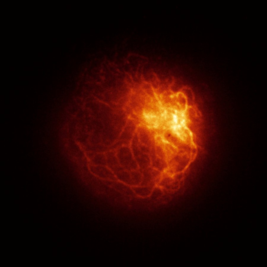
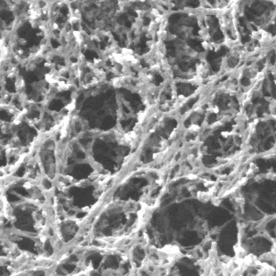
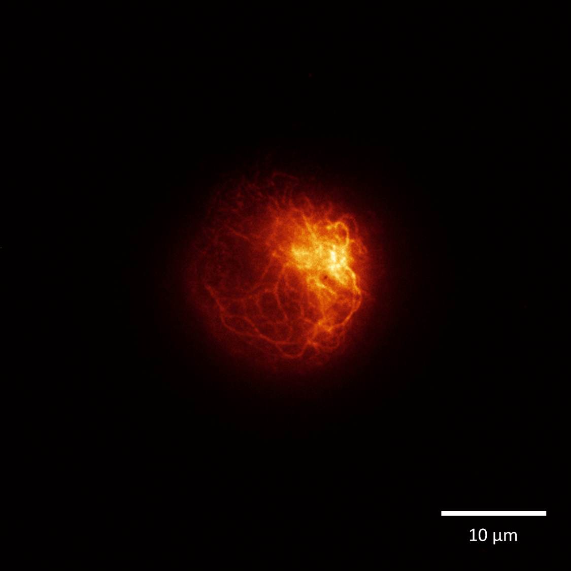
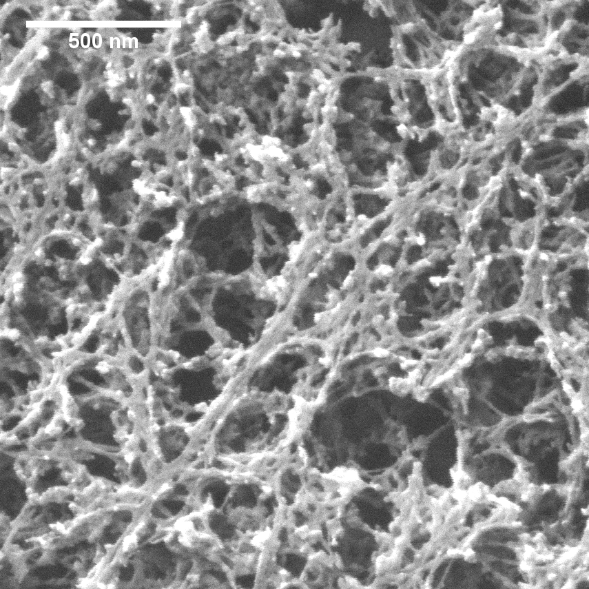
The actin cortex
What is the role of the actin cortex for particular cellular functions, such as migration? Does the cortex structure, dynamics or mechanics alter during adhesion? After the
application of particular cytoskeletal drugs? Or with the composition of the cytoskeletal elements in general?
In order to answer such questions, we are using fluorescent recovery after photobleaching, super resolution and electron microscopy as well as atomic force microscopy.
This project is embedded in the SFB 1027.
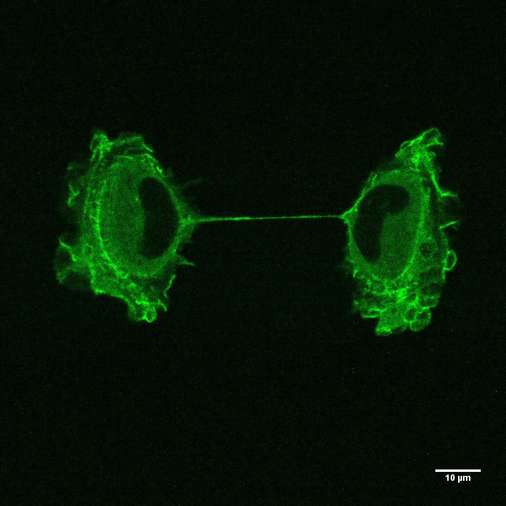
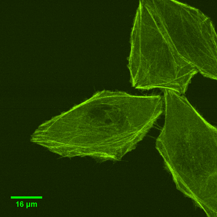
Cell mechanics
Mechanical properties of cells have a high impact on cellular functions, such as the capacity to invade tissue or to migrate. We are testing the impact of
cytoskeletal elements on mechanical properties, e.g. the effect of vimentin in immune cells or the presence of microtubules in microtentancles. Furthermore,
we are interested in potential alterations of mechanical properties due to nanoparticles or particular chemical substances. For measuring cell mechanics, we are
using different techniques depending on the state of adhesion of the cells, e.g. AFM, microfluidic devices, or rheometer.
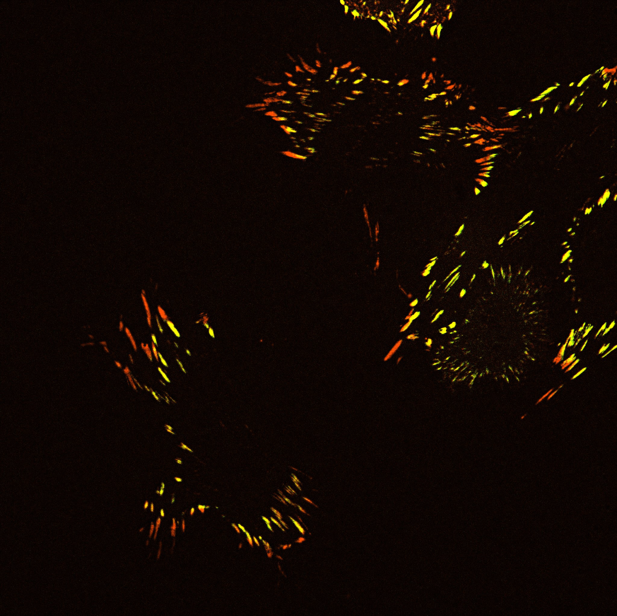
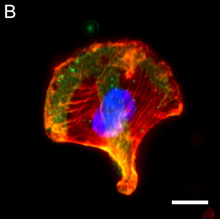
Polarity
How does the shape of a cell changes from a symmetric, non-polar form to a polar one with a well-defined front and back? Which role do cytoskeletal elements play in the
establishment of polarity? And how can we alter this symmetry breaking? We are using micropatterns and fluorescence microscopy to answer such questions.